Neuroplasticity, Culture and Society
Introduction1
The most fundamental difference between the human brain and those of other mammals is the greater extent to which development of its structure and function is influenced by sensory input. This sensitivity to the environment rests on four features common to all mammals and one unique to human beings. First, neurocognitive capacity increases across the phylogenetic hierarchy primarily through increases in the overall number of brain cells and their interconnections. Second, cells require sensory input from the environment to maintain their vitality and functionality. Third, cognitive functions such as perception, memory and thinking arise from the integrated activity of multi-neuronal systems involving multiple brain areas and are not properties of a specific anatomical location dedicated to a specific cognitive operation. Fourth, neurons activated by sensory input develop connections with other neurons and thus constitute the multi-neuronal systems; environmentally induced activity thus shapes both the structure and function of the brain. Fifth, only human beings shape the environments that in turn shape their brains.
Neuroplasticity refers to change in neural structures and is usually the result of activity-dependent change in the interconnections among cells that constitute the structures. Enduring changes in structure result from repeated activation of some cells and pathways more than others, following the principle that neurons that fire together wire together. The neuroplastic potential of the human brain is greater than that of our nearest primate cousins due to changes in two parameter settings in pre and post natal neurodevelopment. One is a further increase in the number of neurons. The second is increased length of time after birth during which interconnections among neurons are easily shaped by environmental input. These two changes make it possible for environmental input to create more elaborate and powerful neural functional structures. The changes in parameter settings are the result of Darwinian biological evolution, but together with the fact that humans alter the environment that provides the formative sensory stimulation, they provide the foundation for cultural evolution.
Cultural evolution differs from Darwinian biological evolution in several important ways. Cultural evolution creates more rapid, more incremental, and more widespread population variability. Cultural and biological evolution also differ in the way information is stored so as to provide continuing influence on function. In biological evolution, information is stored in the largely stable base sequence of DNA molecules. In cultural evolution, the information is stored in the minds and behavior of adult members of society; in cultural artifacts such as books, architecture, and works of art; and in social institutions including laws, customs, and schools. In biological evolution the information is stored in identical and complete form in many individuals. In cultural evolution, the information is distributed in different and incomplete form across many individuals and artifacts.
The extent of our neuroplasticity, and our associated ability to alter the ways our minds and brains work by altering the environment that shapes them, has only recently become known to human beings. Attention to the implications of this for ethics is even more recent. Indeed, Judeo-Christian influences on contemporary Western thinking about morality and ethics have included the view that all aspects of human biology, including our minds and brains, were designed or created more or less in their adult form by the active hand of an external almighty. Both codes for ethical behavior and human characteristics and capabilities were largely given to us by a power external to us and much more powerful than we are. Debates about good and evil, and human responsibility and free-will have taken place within this conceptual field. The first major challenge to this posed by biological science was Darwin’s theory of evolution. It was hotly contested by church authorities when first introduced, and continues to be hotly contested by some religious communities today. The fact that human nature has arisen from single cell organisms through random mutation and selective pressures from an environment that is itself also contingent, reframes the discussion about ethics, accountability, responsibility, good and evil. In this context, some have gone so far as to suggest that ethical behavior, and most important aspects of human thought, have evolved through these same Darwinian processes, although such views are increasingly at odds with current thinking about brain functional organization and their proponents are unable to ascertain the type of data necessary for real scientific inquiry. The new science of neuroplasticity provides the second major change in the terms of ethical considerations introduced by biological sciences.
Understanding the centrality of neuroplasticity in human brain development, and the power of cultural evolution that rests upon it, provides a new biologically based understanding of the relationship between human beings and the environment. The first phase of that relationship is the aforementioned trans-generational dialectic between human capabilities, human actions, and human-created expectations based on the influence of the human-made environment on neurocognitive development. In this phase, developing individuals have limited ability to act on the environment but are profoundly affected by it. A homology is created between the external environment and internal structures because the brain shapes itself to the recurring features of the specific environment in which it develops. By young adulthood, however, there is a fundamental shift in the relationship between the individual and the environment. The powerful neuroplastic processes in the developing brain are replaced by the less powerful ones of adulthood, and now established internal structures are self-maintaining. Individuals are now able to act on the environment and do so to make the environment match established internal structures. They feel and function better when there is a match between internal and external. As a result, there is a “neurobiological antagonism to difference.”2 These processes are central to what it means to be a human being rather than any other animal on earth, and they frame the discussion of human responsibility and free will in a different way than it was before and after application of Darwinian insights to considerations of ethics.
This chapter will review the neuroscience of neuroplasticity in human beings and other mammals. It will present a contemporary neural systems view of brain functional organization, review evidence on the importance of sensory input to maintain neuronal viability, and describe studies that demonstrate that the nature of that input influences brain structure and function.
The centrality of socially generated stimulation will be discussed through citation of the work of Harlow and Mears with infant monkeys, more recent work in rats identifying epigenetic changes in DNA structure leading to life long effects of early maternal behaviors, and the work of the Russian developmental psychologist Vygotsky and early 20th century psychoanalytic ego-psychologists on the role of interpersonal interactions in creating internal mental structures. The next section will present brain-imaging studies that have demonstrated the effects of environmentally induced activity on human brain structure and function. The final section will describe some of the ways established internal structures act on the environment to make it match those structures. The overall goal is to lay the groundwork for a broad-ranging consideration of the issues raised and their implications for politics and ethics.
Part One: A Contemporary Neural Systems View of Human Brain Function
There are 100 billion neurons in the human brain each directly connected to over 1,000 other neurons. Consistent with this massive interconnectivity, learning simple associations between stimuli leads to altered responses in millions of cells distributed across wide expanses of cortical territory.3 When people perform simple cognitive operations, multiple brain areas in both cerebral hemispheres become more active, and others decrease their activity.4 Moreover, when even simple tasks are repeated minutes or hours later, there is a different pattern of task-related regional activation changes, with5 or without6 deliberate efforts to teach or learn the tasks. In real life, most of the things we do we have done before, so that the brain activations associated with them have been different at different times. As people get older, there are also common changes across individuals, so that the same tasks are done by different combinations of brain areas at different ages.7 Furthermore, if the same component cognitive operation is performed as part of different overall cognitive functions, the pattern of regional brain activation associated with that component operation is different.8
These relatively recent observations are consistent with the notion of cerebral functional systems described by the early twentieth-century Russian neuropsychologist A.R. Luria.9 Luria noted that localized injuries rarely affected only one cognitive operation, but usually affected multiple. He also noted that individual cognitive operations were affected by injuries in multiple different areas of the brain. He concluded that while groups of cells in a specific anatomic location might collectively have some elementary tissue function, such functions do not correspond to mental functions like perception, memory or cognition. Mental operations are instead properties of multi-component functional systems. Like other systems, cerebral functional systems perform constant functions through means or components that vary from instance to instance. Functions are properties of a system and not of a specific anatomic location. Most contemporary views of the functional organization of the human brain are based on such systems.
This modern view contrasts with nineteenth-century concepts of phrenology and related twentieth-century concepts of modularity.10 Phrenology and modularity posit that specific cognitive operations are performed at circumscribed, localized anatomic sites, and that the function of these sites is the specific cognitive operation. In contrast, the twenty-first century systems view posits that ensembles of cells at different locations have different characteristics like different letters of an alphabet. Cognitive functions emerge from combinations of different local units just as words emerge from combinations of letters. In addition, there may be a few localized units in the systems model that are also stand alone modules for simple cognitive operations; perhaps operations that evolved prior to the primate, or even the mammalian line. Like the single letter words “a” and “I”, such modules could serve both as free standing cognitive modules and as components of larger systems.
The dynamic systems view helps explain the striking fact that when one hemisphere of the brain must be surgically removed in very young infants, their subsequent cognitive development is largely normal and all cognitive operations are performed with the remaining hemisphere.11 Even when the left or language hemisphere is removed, near-normal language function is supported by the right hemisphere. As with the other examples of developmental neuroplasticity discussed below, these relocations and reconfigurations of brain functional architecture are more easily understood in the systems/emergent property view than in the phrenology/modularity view.
Part Two: Sensory Stimulation and Neuronal Viability and Growth
The brain requires sensory stimulation to maintain structural integrity. Information-processing structures along afferent pathways from peripheral sensory receptors to cortical processing centers atrophy without sensory input. The number of ganglion cells in the retina that carry excitation from photoreceptor cells in the eye to the first relay station in the brain, is decreased to 10% of normal in dark reared chimpanzees;12 after dark-rearing, cats and rats have smaller than normal ganglion cells;13 and rod and cone photoreceptor cells in the eyes of chicks are morphologically abnormal after four weeks with opaque coverings of the eye.14 Both the number and size of cells are reduced by as much as 30-40% in the lateral geniculate of cats and monkeys deprived of visual input during the initial weeks of life.15 The effects continue along the information input pathway to the visual cortex where the number, size, and density of connections among cells are decreased and the organization of cells is altered.16 Studies of olfactory deprivation have yielded a similar picture.17 Effects of sensory deprivation on structural integrity can be decreased by injection of nerve growth factor into the cerebral spinal fluid within the brain during the period of deprivation.18 This naturally occurring substance is produced and released by cells stimulated by sensory input, thus providing further evidence of the association between neuronal activity and neuronal viability and growth.19
Effects of sensory deprivation on development of brain functional organization follow from these effects on cell viability and growth. Neurons at each stage of processing compete for connections with neurons at each subsequent stage, with neurons that fire more often gaining territory. These effects were investigated systematically in Hubel and Wiesel’s Nobel Prize-winning studies of kittens and monkeys.20 Recording electrical activity from hundreds of cells in the area of the brain that receives visual information, they determined that in animals raised under normal conditions most cells respond to inputs from both eyes (approximately 85% in the kitten, 65% in the monkey). Many of these responded somewhat more frequently to input from one eye, with such eye preferences divided evenly between the eyes. Similarly, of the monocularly responsive cells, half responded exclusively to the right eye and half to the left. However, when an eye was sutured closed shortly after birth and then reopened 10 weeks later, 85% or more of cells responded preferentially to the previously non-deprived eye, and few if any cells responded exclusively to the previously deprived eye. Responses to stimulation of the previously deprived eye were slow to start, decreased in amplitude and easily fatigued when present at all.
Hubel and Wiesel also demonstrated two additional features of the effects of sensory-induced neuronal activity on the development of brain structure and function that are of particular relevance to cultural evolution. When visual input to the deprived eye is restored, the altered pattern of cortical cell sensitivities persists despite the fact that both eyes are now receiving unobstructed visual input. As long as neurons from the previously non-deprived eye remain active, they are able to maintain their abnormally acquired hegemony. If, however, the previously non-deprived eye is occluded while the animal is still young enough, the abnormal response pattern can be normalized or reversed in favor of the previously deprived eye.21 The first conclusion of particular interest in relation to cultural evolution is that socially generated activity can create unusual structures that alter the interaction with the environment so as to maintain themselves. In this case, when the eye was occluded, cortical structure changed so as to be unusually responsive to input from only one eye. When the occlusion was removed and input was available to both eyes, the brain still registered input almost exclusively from only one eye. The neural resources necessary to process input now available from the previously occluded eye were absent. They had been appropriated by the active eye during the period of unilateral occlusion, and the active eye maintained the extra resources because it kept those resources actively engaged in processing input within the systems that had appropriated them. This situation could be reversed by occluding the previously open eye, demonstrating that the plastic potential remained, that the brain could be shaped or normalized by corrective intervention, and that without such active intervention the normal pattern could not reassert itself even in a normal situation. The second conclusion of particular interest is that active intervention to normalize or reverse the effects of the initial unilateral occlusion was only effective in young animals. After a certain stage in development, often referred to as the critical period, there is a higher degree of stability in established neural structures, in part because neurochemical mechanisms that support neuroplasticity are less powerful in older individuals.
In further work, Hubel and Wiesel demonstrated that altering the nature or content of the visual stimuli changes the functional organization of the visual cortex even when the stimuli are viewed normally by both eyes. For example, some cells in the visual cortex respond selectively to moving objects, with each cell having maximum sensitivity to movement in a particular direction. Other cells respond selectively to lines (i.e., object edges), with each of these cells having maximum sensitivity to lines of a particular orientation. Kittens raised in strobe light that prevents appreciation of movement have decreased numbers of motion sensitive cells.22 Presumably cells that would have been specialized for movement detection became selectively responsive to some other aspect of visual information instead. Kittens raised in dark except for exposure to stripes moving from left to right have a marked increase in the proportion of cells selectively responsive to left/right rather than right/left movement.23 Similarly, kittens exposed to vertical black and white stripes for a few hours each day, but otherwise reared in darkness, have cortical cells with vertical line orientation preferences, but none with preferences for other orientations.24 Kittens raised wearing goggles that allowed them to see only vertical lines in one eye and horizontal lines in the other, have fewer than the normal number of cells that respond to oblique lines. Moreover, cells responsive to vertical lines are active only with stimulation of the eye that had been exposed to vertical lines, and cells responsive to horizontal lines are active only with stimulation of the eye that had been exposed to horizontal lines.25
The extent of neuroplastic potential in the developing mammalian brain is remarkable. In adult rats that had an eye removed at birth, stimulation of their whiskers led to electrophysiological and metabolic activity within the visual cortex.26 Apparently neurons in what is normally a visual processing area came instead to respond to input from the whiskers when deprived of input from the eye. In perhaps the most dramatic demonstration of plasticity, the optic nerve in one-day-old ferrets was rerouted to provide visual rather than auditory input to what is normally the auditory cortex. The auditory cortex developed a functional organization of ocular dominance columns highly similar to the normal visual cortex rather than its usual tonotopic structure, and the ferrets saw with what would normally have been the auditory regions of the brain.27
The studiessummarized in thissection provide evidence that mammalian brains(and minds) develop concrete perceptual structures, capabilities, and sensitivities based on prominent features of the rearing environment, and then are more able and more likely to see those features in the sensory mix of new environments encountered subsequently. Or to turn it around, mammals have limited ability to see even prominent features of a new environment if those features were absent from their rearing environment.
Part Three: Social Interactions as the Source of Early Environmental Stimulation
The class mammalia is named on the basis of the presence of mammary glands. It is defined on the basis of nourishing young with milk and a series of physical features including a chain of small ear bones, four optic lobes in the brain, a particular mandibular structure, a muscular diaphragm separating the lungs and heart from the abdomen, only a left aortic arch, warm blood with red blood cells lacking nuclei, and viviparous reproduction. In studies of infant monkeys and wire mesh surrogate mothers, Harlow and Mears provided a radical correction to this definition, adding another central feature that in many ways is more important than all the others. Infant monkeys were separated from their mothers and raised in cages with access to both a wire mesh and a cloth surrogate mother. Both surrogate mothers were kept at the same temperature as normal monkey mothers. One-half of the monkeys received milk from the wire mesh mother and one-half from the cloth mother. Both groups spent much more time on the cloth than the wire mesh mother. The differential was greater by only a small amount when the cloth mother was the source of milk. The preference for the cloth mother became greater over time in both groups, the opposite of what would be expected from a food/hunger reduction conditioning model which would predict increasing preference over time for the food-providing surrogate mother. Harlow and Mears concluded that “the disparity [in favor of selecting the cloth mother independent of which mother provides milk] is so great as to suggest that the primary function of nursing as an affectional variable is that of ensuring frequent and intimate body contact of the infant with the mother.”28 In other words, instead of the provision of milk being the end goal of mother infant interaction in and of itself, it is a means of ensuring contact between the mother and the infant because this contact is essential for provision of sensory stimulation necessary for brain development, and for production of population variability through variability in that stimulation.
Real living mothers and other parenting figures vary in the ways they stimulate their infants and children. Naturally occurring differences in these parenting behaviors have life long and specific effects on the brains and behavior of their offspring, and changes in DNA structure that mediate these effects have been identified in studies of rats.29 Mother rats differ in the amount of time they spend licking and grooming their pups, and the in the ways they position themselves for nursing. Michael Meaney and colleagues found that adult rats that had been licked more as pups had decreased behavioral and hormonal responses to stress, and greater spatial learning abilities—a capacity in which areas of the hippocampus play an important role.30
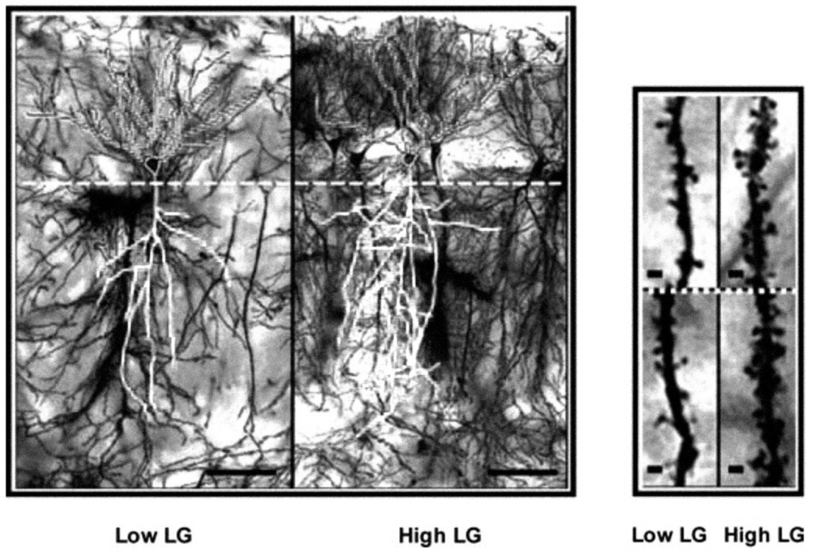
Examining brain chemistry and structure, they found greater levels of specific types of messenger RNA that carry the information from the DNA to parts of the cells that synthesize the glucocorticoid receptors important in regulating stress responses and the NMDA receptors important in promoting neuroplasticity. Direct examination of the hippocampus revealed that offspring of high licking mothers had longer neurons with more branches and interconnections (FIG.1).31 Direct examination of the DNA identified actual changes in the genes associated with stress response as a result of the degree of maternal licking. Shortly after birth, the surface of DNA is largely covered by small chemical complexes called methyl groups. These methyl groups limit access to the DNA and thereby limit activation or expression of genes. Experiences during the first weeks of life can lead to selective removal of these methyl groups, making some genes more active. The effects of experience on methylation are much greater during the first three weeks of a rat’s life than thereafter, and changes induced by experience during this critical period usually remain relatively unchanged throughout the rat’s adult life. Maternal licking initiates a series of neurochemical processes that selectively demethylate genes that produce the glucocorticoid receptors in the hippocampus and frontal lobes that turn off the stress response.
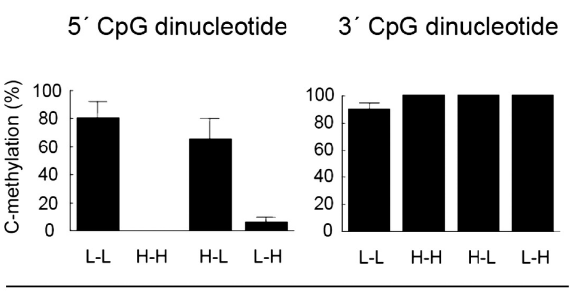
To ensure that these observations were due to the differences in maternal behavior, and not to genes that high licking mothers passed on to their offspring, Meaney and colleagues had pups born to low licking mothers raised from birth by high licking mothers, and vice-versa.32 When these rats became adults, their stress responses and the methylation of their DNA (FIG. 2)33 were both consistent with the type of mother that reared them and not with the type of their biological mother.
Two other aspects of this work are also of relevance to cultural evolution. First, when given learning tests in high stress environments, adult rats raised by low licking mothers out performed rats raised by high licking mothers. This demonstrates the adaptive value of the population variability induced by cultural evolution. Second, some of the persistent neurochemical and behavioral effects of maternal care of female infants affect the way the infant functions as a mother herself when she becomes an adult. Females that had been separated from their mothers when they were infants, showed lower than normal gene expression in areas of the brain associated with maternal behaviors when they themselves became mothers.34
They also licked and crouched over their pups less often than other mothers,35 and their generally decreased ability to maintain attention and increased response to stress have been hypothesized to further compromise their maternal competence.36 Such intergenerational effects are potentially self-propagating and even self-amplifying. Moreover, since litter size37 and food availability38 can influence the amount of licking and other behavioral interactions between mother and infant, a variety of environmental factors can influence maternal behaviors and their impact, across generations, on a range of individual and group behaviors. All this depends on the post-natal sensitivity of the mammalian brain to sensory stimulation, and the proximity of mammalian infants and mothers ensured by nursing.
Part Four: The Human Rearing Environment
Human rearing behaviors are more complex and more varied than those of other mammals, and include massive social components and influences from extended families, communities, and nation states. The extra-familial influences include schools, mass media, arts, laws, and customs. The human social and economic environments also affect the states of mind, time, and energy of the parents, thus affecting their interactions with their offspring in a manner analogous to the effects of food supply on rat maternal behavior. And although beyond the scope of this chapter to discuss, the huge role of language—spoken and written—in facilitating the influence of the human-made environment on the development of children must be noted, along with the fact that the latter is itself clearly a product of cultural evolution and it seems increasingly probable that the former is in large part as well.
At birth, human infants can distinguish their mother’s language from other languages based on stimulation received in utero.39 Within hours of birth they show a selective interest in looking at the human face, with the interest greatest for the full face as experienced in social interactions rather than for the face in profile. Within days they prefer their mother’s face and voice to those of others.40 Within this context, parents provide objects of play and structure interactions and activities. As Kenneth Kaye has remarked, “social interference in the object-directed activities of babies is such a commonplace occurrence that few authors have remarked on its absolute uniqueness to our own species.”41 The brains and minds of human infants and children develop while closely linked to the minds and brains of their biobehaviorally mature caregivers. The characteristics of the adults shape the stimulation that shapes the growing brains of the children through the small details and general rhythms of the child’s experiences. The child integrates input from progressively larger circles of direct interaction, beginning with primary care givers and growing to include extended family members and then members of the community and society more broadly.
While some of the social input is actively shaped and provided by others, much is just absorbed through essentially constant imitation. Within two days of birth, infants will stick out their tongues and move their heads in imitation of an adult doing so.42 From infancy on, children learn how to do things simply by watching them done. They imitate the goals of action even by different means and imitate a parent’s affective response to new stimuli.43 Mirror neurons fire when people (and monkeys) watch an act being done, and many times these same neurons are then active when the individual performs the action previously observed.44 Similarly, looking at someone else in pain activates the same regions of the brain as are active when the observer experiences pain him or herself.45
The earlier cited work of Hubel and Wiesel demonstrated that environmentally induced neuronal activity shaped the development of cerebral functional structures, following the principle that neurons that fire together wire together. In human development, active parental and community interventions and nearly constant imitation of what is seen and heard produce intensive and repetitive firing of neuronal ensembles and circuits. This environment-induced neural activation shapes brain development to be consistent with the largely human-made rearing environment.
Well before the relevant neuroscience research, psychologists were aware of the role of the social environment in shaping mental development, describing the processes in language remarkably similar to what would be suggested by the subsequent work of Hubel, Wiesel, Meaney and others. Writing in 1926, Fenichel states that “changes in the ego, in which characteristics which were previously perceived in an object [usually an important person] are acquired by the perceiver of them, have long since been familiar to psychoanalysis.”46 Freud described identification as “the assimilation of one ego to another one, as a result of which the first ego behaves like the second in certain respects, imitates it and in a sense takes it up into itself.”47 Greenson stated that “identification with an object means that… a transformation of the self has occurred whereby the self has become similar to the external object… one can observe behavior, attitudes, feelings, posture, etc., which are now identical to those characteristics belonging to the external object”,48 and that at early stages of development “perception impliestransformation of the self.” Reich explained that “the child simply imitates whatever attracts his attention momentarily in the object… normally these passing identifications develop slowly into permanent ones, into real assimilation of the object’s qualities.”49 Writing from a different cultural and intellectual context, the Russian psychologist Lev Vygotsky described the process: “In the early stages of development the complex psychological function was shared between two persons: the adult triggered the psychological process by naming the object or by pointing to it; the child responded to this signal and picked out the named object either by fixing it with his eye or by holding it with his hand. In the subsequent stages of development… The function which hitherto was shared between two people now becomes a method of internal organization of the psychological process. From an external, socially organized attention develops the child’s voluntary attention, which in this stage is an internal, self-regulating process.”50
Part Five: Brain Imaging Demonstrations of Environment-Induced Brain Organization in Human Beings
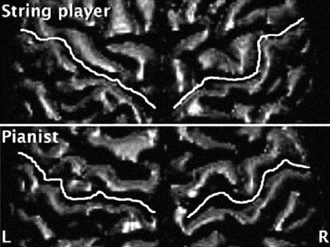
Brain imaging studies have now demonstrated changes in brain structure and function that result from unusual motor activity or sensory input during childhood and persist into adulthood. One set of studies has examined differences in brain structure and function as a result of practicing a musical instrument during childhood. A socially and culturally created and induced activity on multiple levels, intensive practice of string instruments leads to selective increase in volume of the right somatosensory and motor areas associated with the rapid, fine motor movements of the fingers of the left hand that provide intricate and fast moving sequences of pressure to the strings. The changes in the brain are greater in adults who practiced more hours and began practicing at younger ages.51 Figure 3 shows this bulked up motor cortex in the right hemisphere of string players (the increase in volume is actually visible to the naked eye!) and bilaterally in piano players who practice with both hands.efn_note]M. Bangert and G. Schlaug, “Specialization of the specialized in features of external human brain morphology,” in European Journal of Neuroscience, 24 (2006): 1832-4.[/efn_note]
The second set of studies looked at brain activations in the normal visual areas of the brain in adults who were blind at birth or shortly thereafter, or the normal auditory areas of the brain in adults who were deaf at or shortly after birth.
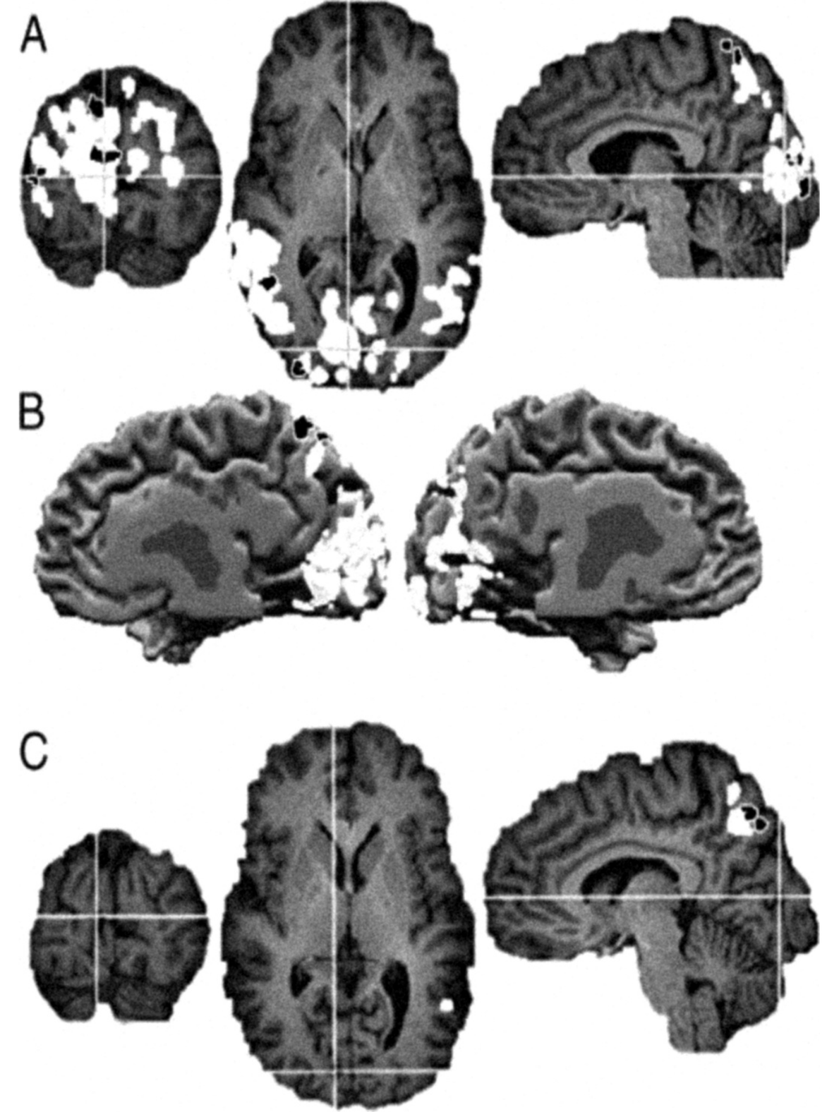
Directly analogous to the selective sensory deprivation experiments of Hubel and Wiesel, the findings were also analogous. In early blind subjects, the area of the brain that is normally the site of early visual processing is activated instead by auditory and tactile stimulation (FIG. 4),52 and is also more active during language processing tasks than is the case in sighted people.53 Apparently, when the normal sensory input to the area was absent, other sensory input and cognitive operations moved into the territory. Moreover, among the blind individuals, memory performance was higher in the individuals who made more use of the “visual” areas during the memory task.
The results of these new imaging studies in humans are what is expected based on the studies in animals, the increased plasticity of the human brain and the very active structuring by human adults of the rearing environment and developmental experiences of their offspring. As mentioned above, a 1994 study showed that if one eye of a rat is removed at birth, stimulation of their whisker when they are adults activates cells in what is usually visual cortex.54 The demonstration of similar activation in the humans who are blind from early life, then, is no surprise. The demonstration of changes in brain morphology as a result of practicing a musical instrument extends things a bit beyond the animal studies in that practicing music is clearly a socially constructed human activity, and it is impressive that the environmentally induced changes can be seen with the naked eye when data from multiple individuals is averaged together. But the demonstration of changes is at a gross anatomic level and does not reveal more fine grained changes in structure and function. These studies, however, are important in the workman-like effort of science to test assumptions and build bridges that link different sets of data. We do not yet have methods and data in people to enable us to demonstrate effects of parental actions on axonal branching in the hippocampus, as Meaney and colleagues have done in rats, but by linking the data and theory from animal studies to human beings with the above cited imaging studies, scientists complete an evidentiary loop and increase confidence in the application to human beings of principles based on the data from animals.
Summary and Conclusions
Functional properties of individual neurons in the human brain differ little from those of individual neurons in the brains of other primates. The large differences in function between the human brain and other primate brains result instead from the increased number of cells and interconnections among them, the extended period after birth during which the brain is highly susceptible to shaping by environmentally induced neuronal activity, and the fact that humans alone alter the environment that produces the neuronal activity that shapes the brains of their offspring. Together these factors constitute neuroplasticity and cultural evolution. Cultural evolution produces changes in human capabilities, desires and expectations much more rapidly and through very different mechanisms than does Darwinian biological evolution. It is a cross generational and social process which shapes individual actions, and these actions then in turn contribute to the social and cross generational influences that shape other individuals.
We humans are not handed a set of fixed capabilities, developed desires and inclinations, and standards for ethical conduct. All three of these critical aspects of human being are in dynamic interplay through human history; such is our neurobiological relationship with our natural and human-made environments. Our ability to shape our environments, and through that to shape our minds, brains, and behavior, begets complex responsibility, promising opportunity and political contestation. While political process may have been seen as contests between communities of established interests and positions, or between candidates each attempting to persuade people they are the candidate that best represents their interests and opinions, it now must be seen as fundamental in creating the positions it seeks to represent or adjudicate. When we turn for guidance to ethical traditions and principles, we must understand that there is as much tradition as principle. Solid ground is difficult to identify in the dynamic interplay of socially con- structed or cultured brains, and the human-made culture in which our brains develop, live and function.55
Marxist and post-Marxist theory has been clear that human constructed, and historical rather than natural, social and economic structure and process heavily influence the way people understand their own self-interest and think more broadly. But while this powerful perspective provides precedent and vocabulary for approaching the interplay between brain and culture, it requires further consideration of the more radical reformulations of neuro-plastic science.56 Organized religion has had deep disagreement with Marxism in the past, as it has had with evolution science. Implications of new neuroscience challenge the scope and assumptions of both theology and existing social theory. What directs the course of human development? What is better and what is worse? Are there criteria for judgment? Can the transgenerational and multifactorial processes be rationally and consensually directed?
- Editors’ Note: Another version of this article, titled “Shaping the Environments that Shape Our Brains: A Long Term Perspective”, was published in Deborah Hauptmann and Warren Neidich, Cognitive Architecture: From Biopolitics to Noopolitics: Architecture and Mind in the Age of Communication and Information (Rotterdam: 010 Publishers, 2010).
- Bruce E. Wexler, Brain and Culture: Neurobiology, Ideology and Social Change (Cambridge, MA: MIT Press, 2006), 212.
- E.R. John, Y. Tang, A.B. Brill, et al., “Double-labeled metabolic
maps of memory,” in Science, 233 (1986): 1167-75. - M. D’Esposito, “From cognitive to neural models of working memory,” in Philosophical Transactions of the Royal Society of London – Series B: Biological Sciences, 362 (2007): 761-72.
- R.A. Poldrack, V. Prabhakaran, C.A. Seger, C.A., et al., “Striatal activation during acquisition of a cognitive skill,” in Neuropsychology, 13 (1999): 564-74.
- C. Kelly, J.J. Foxe, and H. Garavan, “Patterns of normal human brain plasticity after practice and their implications for neurorehabilitation,” in Archives of Physical Medicine and Rehabilitation, 87 (2006): S20-9; I. Loubinoux, C. Carel, F. Alary, et al., “Within-session and between-session reproducibility of cerebal sensorimotor activation: a test-retest effect evidenced with functional magnetic resonance imaging,” in Journal of Cerebral Blood Flow and Metabolism, 21 (2001): 595-607.
- W.D. Gaillard, L. Hertz-Pannier, S.H. Mott, et al., “Functional anatomy of cognitive development: fMRI of verbal fluency in children and adults,” in Neurology, 54 (2000): 180; G.T. Stebbins, M.C. Carrillo, J. Dorfman, et al. “Aging effects on memory encoding in the frontal lobes,” in Psychology and Aging, 17 (2002): 44-55.
- K.J. Friston, C.J. Price, P. Fletcher, et al., “The trouble with cognitive subtraction,” in NeuroImage, 4 (1996): 97-104.; B.E. Wexler, “Using fMRI to study the mind and brain,” in R. Shulman and D. Rothman, eds., Brain Energetics and Neuronal Activity (West Sussex: John Wiley and Sons, 2004), 279-94.
- A.R. Luria, The Working Brain, trans. B. Haugh, (New York: Basic Books, 1973); L.S. Vygotsky, Mind in Society: The Development of Higher Psychological Processes (Cambridge, MA: Harvard University Press, 1978)
- (see Wexler, 2004, 2006)
- J.A. Ogden, “Phonological dyslexia and phonological dysgraphia following left and right hemispherectomy,” in Neuropsychologia, 34 (1996): 905-18; R. Werth, “Visual functions without the occipital lobe or after cerebral hemispherectomy in infancy,” in European Journal of Neuroscience, 24 (2006): 2932-44.
- E. Rasch, H. Swift, A.H. Riesen, et al., “Altered structure and composition of retinal cells in dark eared animals,” in Experimental Cell Research, 25 (1961): 348-63.
- Ibid.
- H. Liang, D.P. Crewther, S.G. Crewther, et al., “A role for photoreceptor outer segments in the induction of deprivation myopia,” in Vision Research, 35 (1995): 1217-25.
- (e.g., D.H. Hubel, “Deprivation and development,” in Eye, Brain and Vision (New York: Scientific American Library, 1988), 191-217; D.H. Hubel and T.N. Wiesel, “The period of susceptibility to the physiological effects of unilateral eye closure in kittens,” in Journal of Physiology, 206 (1970): 419-36; T.N. Wiesel and D.H. Hubel, “Effects of visual deprivation on morphology and physiology of cells in the cat’s lateral geniculate body,” in Journal of Neurophysiology, 26 (1963): 978-93; C. Kupfer and P. Palmer, “Lateral geniculate nucleus: histological and cytochemical changes following afferent denervation and visual deprivation,” in Experimental Neurology, 9 (1964): 400-9; S.M. Sherman, K.P. Hoffman, and J. Stone, “Loss of a specific cell type from dorsal lateral geniculate nucleus in visually deprived cats,” in Journal of Neurophysiology, 35 (1972): 532-41; S.M. Sherman and K.J. Sanderson, “Binocular interaction on cells of the dorsal lateral geniculate nucleus of visually deprived cats,” in Brain Research, 37 (1972): 126-31; M. Tigges, and J. Tigges, “Parvalbumin immunoreactivity in the lateral geniculate nucleus of rhesus monkeys raised under monocular and binocular deprivation conditions,” in Visual Neuroscience, 10 (1993): 1043-53.
- e.g., G.K. Aghajanian, and F.E. Bloom, “The formation of synaptic junctions in developing rat brain: a quantitative electron microscopic study,” in Brain Research, 6 (1967): 716-27; B.G. Cragg, “What is the signal for chromatolysis?” in Brain Research, 23 (1970): 1-21; E. Fifková, “Changes of axosomatic synapses in the visual cortex of monocularly deprived rats,” in Journal of Neurobiology, 2 (1970): 61-71; A. Kumar, and R. Schliebs, “Postnatal laminar development of cholinergic receptors, protein kinase C and dihydropyridine-sensitive calcium antagonist binding in rat visual cortex. Effect of visual deprivation,” in International Journal of Developmental Neuroscience, 10 (1992): 491-504; A. Kumar, and Schliebs, R., “Postnatal ontogeny of GABAA and benzodiazepine receptors in individual layers of rat visual cortex and the effect of visual deprivation,” in Neurochemistry International, 23 (1993): 99-106; P. Rakic, I. Suner, and R.W. Williams, “A novel cytoarchitectonic area induced experimentally within the primate visual cortex,” in Proceedings of the National Academy of Sciences of the United States of America, 88 (1991): 2083-7; S. Robner, A. Kumar, W. Kues, et al., “Differential laminar expression of AMPA receptor genes in the developing rat visual cortex using in situ hybridization histochemistry: Effect of visual deprivation,” in International Journal of Developmental Neuroscience, 11 (1993): 411-24.
- T.E. Benson, D.K. Ryugo, and J.W. Hinds, “Effects of sensory deprivation on the developing mouse olfactory system: a light and electron microscopic, morphometric analysis,” in Journal of Neuroscience, 4 (1984): 638-53; J. Najbauer, and M. Leon, “Olfactory experience modulated apoptosis in the developing olfactory bulb,” in Brain Research, 674 (1995): 245-51; L.C. Skeen, B.R. Due, and F.E. Douglas, “Neonatal sensory deprivation reduces tufted cell number in mouse olfactory bulbs,” in Neuroscience Letters, 63 (1986): 5-10.
- N. Berardi, A. Cattaneo, A. Cellerino, et al.,“Monoclonal antibodies to nerve growth factor (NGF) affects the postnatal development of the rat geniculocorticalsystem,” in Journal of Physiology-London, 452 (1992): 293; N. Berardi, L. Domenici, V. Parisi, et al., “Monocular deprivation effects in the rat visual cortex and lateral geniculate nucleus are prevented by nerve growth factor (NGF). I. Visual cortex,” in Proceedings of the Royal Society of London, B251 (1993): 17-23; G. Carmignoto, R.Canella, P. Candeo, et al., “Effects of nerve growth factor on neuronal plasticity of the kitten visual cortex,” in Journal of Physiology-London, 464 (1993): 343-60; L. Domenici, A. Cellerino, and L. Maffei, “Monocular deprivation effects in the rat visual cortex and lateral geniculate nucleus are prevented by nerve growth factor (NGF). II. Lateral geniculate nucleus,” in Proceedings of the Royal Society of London, B251 (1993): 25-31; T. Pizzorusso, M. Fagiolini, M. Fabris, et al., “Schwann cells transplanted in the lateral ventricles prevent the functional and anatomical effects of monocular deprivation in the rat,” in Proceedings of the National Academy of Sciences of the United States of America, 91 (1994): 2572-6.
- L. Domenici, A. Cellerino, and L. Maffei, “Monocular deprivation effects in the rat visual cortex and lateral geniculate nucleus are prevented by nerve growth factor (NGF). II. Lateral geniculate nucleus,” in Proceedings of the Royal Society of London, B251 (1993): 25-31.
- Hubel, “Deprivation and development,” 191-217.
- Hubel, “Deprivation and development,” 191-217.
- M. Cynader, N. Berman, and A. Hein, “Cats reared in stroboscopic illumination: effects on receptive fields in visual cortex,” in Proceedings of the National Academy of Sciences of the United States of America, 70 (1973): 1353-4; M. Cynader, and G. Chernenko, “Abolition of direction selectivity in the visual cortex of the cat,” in Science, 193 (1976): 504-5.
- F. Tretter, M. Cynader, and W. Singer, “Modification of direction selectivity of neurons in the visual cortex of kittens,” in Brain Research, 84 (1975): 143-9.
- C. Blakemore, and G.F. Cooper, “Development of the brain depends on visual experience,” in Nature, 228 (1970): 477-8.
- H.B. Hirsch, and D. Spinelli, “Visual experience modifies distribution of horizontally and vertically oriented receptive fields in cats,” in Science, 168 (1970): 869-71.
- J. Toldi, I. Rojik, and O. Feher, “Neonatal monocular enucleation-induced cross-modal effects observed in the cortex of adult rat,” in Neuroscience, 62 (October 1994): 105-14.
- J. Sharma, A. Angelucci, and M. Sur, “Induction of visual orientation modules in auditory cortex,” in Nature, 404 (2000): 841-7.
- Harlow, H.F. and Mears, C., The Human Model: Primate Perspectives (Washington: V. H. Winston & Sons, 1979), 108.
- I.C.G. Weaver, N. Cervoni, F.A. Champagne, F.A., et al.,“Epigenetic programming by maternal behavior,” in Nature Neuroscience, 7 (2004): 847-54; I.C.G. Weaver, J. Diorio, J.R. Seckl, et al., “Early environmental regulation of hippocampal glucocorticiod receptor gene expression: characterization of intracellular mediators and potential genomic sites,” in Annals of the New York Academy of Sciences, 1024 (2004): 182-212.
- Weaver, et al., “Early environmental regulation of hippocampal glucocorticiod receptor gene expression,” 182-212.
- D.L. Champagne, R.C. Bagot, F. van Hasselt, F., et al.,“Maternal care and hippocampal plasticity: evidence for experience-dependent structural plasticity, altered synaptic functioning, and differential responsiveness to glucocorticoids and stress,” in Journal of Neuroscience, 28 (2008): 6037-45.
- I.C.G. Weaver, N. Cervoni, F.A. Champagne, et al., “Epigenetic programming by maternal behavior,” in Nature Neuroscience, 7 (2004): 847-54; I.C.G. Weaver, J. Diorio, J.R. Seckl, M. Szyf, et al., “Early environmental regulation of hippocampal glucocorticiod receptor gene expression: characterization of intracellular mediators and potential genomic sites,” in Annals of the New York Academy of Sciences, 1024 (2004): 182-212.
- Weaver, et al.,“Epigenetic programming by maternal behavior,” 847-54.
- A.S. Fleming, G.W. Kraemer, A. Gonzalez, et al.,“Mothering begets mothering: the transmission of behavior and its neurobiology across generations,” in Pharmacology Biochemistry and Behavior,73(2002): 61-75.
- A. Gonzalez, V. Lovic, G.R. Ward, et al., “Intergenerational effects of complete maternal deprivation and replacement stimulation on maternal behavior and emotionality in female rats,” in Developmental Psychobiology, 38 (2001): 11-32.
- Fleming, et al.,“Mothering begets mothering,” 61-75.
- Ibid.; J.E. Jans, and B. Woodside, “Effects of litter age, litter size, and ambient temperature on the milk ejection reflex in lactating rats,” in Developmental Psychobiology, 20 (1987): 333-44.
- D.M. Lyons, H.Afariana,A.F. Schatzberg, et al.,“Experience-dependent asymmetric variation in primate prefrontal morphology,” in Behavioural Brain Research, 136 (2002): 51-9.
- J. Mehler, P. Jusczyk, G. Lambertz, et al., “A precursor of language acquisition in young infants,” in Cognition, 29 (1988): 143-78.
- G. Carpenter, “Mother’s face and the newborn,” in New Scientist, 21 (1974): 742-4; W.P. Fifer and C.M. Moon, “The role of mother’s voice in the organization of brain function in the newborn,” in Acta Paediatrica (Suppl.), 397 (1994): 86-93; C.C. Goren, M. Sarty, and P.Y.K. Wu, “Visual following and pattern discrimination of face-like stimuli by newborn infants,” in Pediatrics, 56 (1975): 544-9; A. MacFarlane, “What a baby knows,” in Human Nature, 1 (1978); J. Mehler, P. Jusczyk, G. Lambertz, et al., “A precursor of language acquisition in young infants,” in Cognition, 29 (1988): 143-78; Mills, M. and Melhursh, E. “Recognition of mother’s voice in early infancy,” in Nature, 252 (1974): 123-4; R. Spitz and K. Wolf, “The smiling response: a contribution to the ontogenesis of social relations,” in Genetic Psychology Monographs, 34 (1946): 57-125.
- K. Kaye, “Organism, apprentice, and person,” in E. Tronick, ed., Social Interchange in Infancy: Affect, Cognition, and Communication, (Baltimore: University Park Press, 1982), 183-96.
- A.N. Meltzoff and M.K. Moore, “Imitation of facial and manual gestures by human neonates,” in Science, 198 (1977): 74-8; A.N. Meltzoff and M.K. Moore, “Imitation in newborn infants: exploring the range of gestures imitated and the underlying mechanisms,” in Developmental Psychology, 25 (1989): 954-62.
- K. Kaye, The Mental and Social Life of Babies: How Parents Create Persons (Chicago: University of Chicago Press, 1982); M. Klinnet, R.N. Emde, P. Butterfield, et al., “Social referencing: the infant’s use of emotional signals from a friendly adult with mother present,” in Developmental Psychology, 22 (1986): 427-32.
- M. Iacoboni, R.P. Woods, M. Brass, et al., “Cortical mechanisms of human imitation,” in Science, 286 (1999): 2526-8; G. Rizzolatti, L. Fadiga, V. Gallese, et al.,“Premotor cortex and the recognition of motor actions,” in Cognitive Brain Research, 3 (1996): 131-41; M.A. Umilta, E. Kohler, V. Gallese, et al.,“I know what you are doing: a neurophysiological study,” in Neuron, 31 (2001): 155-65.
- X. Gu and S. Han, “Attention and reality constraints on the neural processes of empathy for pain,” in NeuroImage, 36 (2007): 256-67; P.L. Jackson, A.N. Meltzoff, and J. Decety, “How do we perceive the pain of others? A window into the neural processes involved in empathy,” in NeuroImage, 24 (2005): 771-9; T. Singer, B. Seymour, J. O’Doherty, et al.,“Empathy for pain involves the affective but not sensory components of pain,” in Science, 303 (2004): 1157-62.
- O. Fenichel, (1926), “Identification,” in G. Pollock, Pivotal Papers on Identification (Madison, CT: International Universities Press, 1993), 57-74.
- S. Freud, (1933), “Excerpt from Lecture XXXI: The dissection of the psychical personality,” in G. Pollock, Pivotal Papers on Identification (Madison, CT: International Universities Press, 1993), 47-52.
- R.R. Greenson, (1954), “The struggle against identification” in G. Pollock, Pivotal Papers on Identification (Madison, CT: International Universities Press, 1993), 159-75.
- A. Reich, (1954), “Early identifications as archaic elements in the superego,” in G. Pollock Pivotal Papers on Identification, (Madison, CT: International Universities Press, 1993), 177-95.
- A.R. Luria, The Working Brain, trans. B. Haugh, (New York: Basic Books, 1973)
- G. Schlaug, “The brain of musicians: a model for structural and functional adaptation,” in Annals of the New York Academy of Sciences, 930 (2001): 281-99.
- K.E. Weaver and A.A. Stevens, “Attention and sensory interactions within the occipital cortex in the early blind: an fMRI study,” in Journal of Cognitive Neuroscience, 19 (December 2007): 315-30.
- A. Amedi, N. Raz, P. Pianka, et al.,“Early ‘visual’ cortex activation correlates with superior verbal memory performance in the blind,” in Nature Neuroscience, 6 (2003): 758-66.
- J. Toldi, I. Rojik, and O. Feher, “Neonatal monocular enucleation-induced cross-modal effects observed in the cortex of adult rat,” in Neuroscience, 62 (October 1994): 105-14.
- W. Neidich, Blow-up Photography, Cinema and the Brain. (DAP/UCR/California Museum of Photography, 2003).
- W. Neidich, “Pointings” in Lost Between the Extensivity/Intensity Exchange, (Eindhoven: Onomatopee 25, 2009), 65-72.